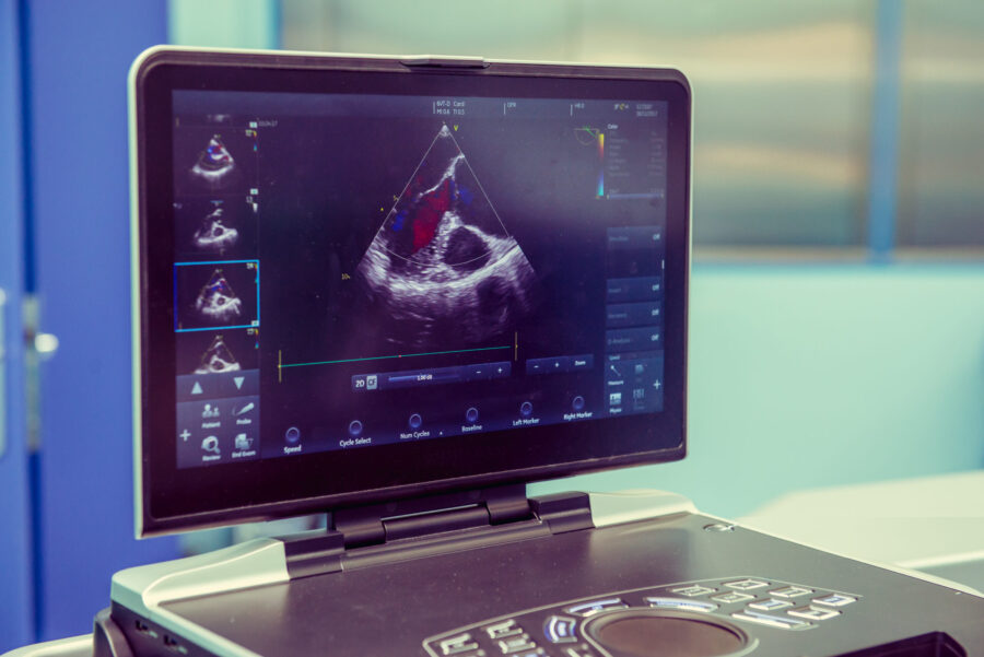
Echocardiography, often referred to as an Echo, is a non-invasive, painless test that uses ultrasound technology to capture images of the heart. This essential tool in cardiology helps doctors diagnose, monitor, and manage heart diseases. In this article, we will delve into the Echo procedure, its types, its significance in modern medicine, and the exciting advancements that offer a glimpse into its future.
Understanding an Echo Procedure:
An Echo uses sound waves to produce images of the heart, showing the size, shape, and movement of the heart’s valves and chambers, as well as the blood flow through your heart. These images provide crucial insights into the heart’s performance, enabling doctors to detect heart conditions, assess heart function, and identify abnormalities.
The test is entirely safe and non-invasive, making it a preferred choice for evaluating heart health. It involves no radiation exposure and poses minimal risk to the patient. The procedure is commonly performed in a hospital or specialized cardiac center and is conducted by a trained echocardiographer.
Why Doctors Recommend an Echo:
- Echocardiography is a versatile diagnostic tool that offers valuable information about the heart. Doctors recommend an Echo for various reasons, including:
- Evaluation of Heart Function: An Echo allows doctors to assess the heart’s pumping ability, the movement of its walls, and the efficiency of blood flow.
- Detection of Heart Conditions: The Echo can identify various heart conditions, such as heart valve abnormalities, heart muscle disorders (cardiomyopathies), congenital heart defects, and pericardial diseases.
- Diagnosis of Heart Diseases: Echocardiography aids in diagnosing coronary artery disease (CAD), heart attacks, and other cardiovascular diseases.
- Monitoring Existing Heart Conditions: Patients with known heart conditions require regular monitoring of their heart function. Echocardiography enables doctors to track disease progression and determine the effectiveness of treatments.
- Guidance for Treatment Plans: Echocardiography provides crucial information to cardiologists, helping them develop personalized treatment plans for patients with heart problems.
Types of Echocardiography:
There are several types of echocardiography, each serving specific purposes. The most common types include:
- Transthoracic Echocardiogram (TTE): This is the most frequently performed Echo procedure. During a TTE, a technician applies a water-based gel to the patient’s chest and places a transducer on the skin. The transducer emits ultrasound waves that bounce off the heart’s structures, creating real-time images on a monitor.
- Transesophageal Echocardiogram (TEE): TEE provides more detailed images of the heart by positioning the transducer closer to the heart. The transducer is attached to the end of a thin, flexible tube (endoscope) that is inserted through the patient’s mouth and gently guided down the esophagus. This procedure is usually performed under light sedation to ensure patient comfort.
- Stress Echocardiogram: A stress echocardiogram combines an Echo with a stress test, which involves exercising on a treadmill or stationary bike. The Echo is conducted both before and immediately after exercise. The test helps evaluate the heart’s response to physical stress, particularly in cases where coronary artery disease is suspected but not evident at rest.
What to Expect During an Echo:
Before the Echo procedure, the patient’s medical history and symptoms are reviewed to ensure that the most appropriate type of echocardiography is selected. The test is typically painless, with minimal discomfort experienced during TEE due to the presence of the endoscope. Patients undergoing a stress echocardiogram will be guided through the exercise protocol according to their physical abilities.
Interpreting the Results:
After the Echo procedure, a qualified cardiologist interprets the collected images. The results provide a wealth of information about the heart’s structure and function, including the size and thickness of the heart walls, the condition of the heart valves, and any abnormal blood flow patterns.
The interpretation of Echo images aids in the diagnosis of various heart conditions and guides treatment decisions. If any abnormalities or heart diseases are detected, the doctor will discuss the findings with the patient and recommend further testing or treatment options as needed.
Advancements in Echocardiography:
As technology continues to evolve, so does echocardiography. Recent advancements have revolutionized the field, enhancing its capabilities and widening its scope. Here are some exciting developments that offer a glimpse into the future of echocardiography:
- 3D Echocardiography: Traditional echocardiography provides two-dimensional images of the heart. However, 3D echocardiography takes it a step further, offering more detailed and realistic views of the heart’s structures. This advancement enables clinicians to accurately assess complex cardiac anatomy, such as congenital heart defects and valvular abnormalities. 3D echocardiography provides valuable information for surgical planning and guides interventional procedures.
- Strain Imaging: Strain imaging is a sophisticated technique that measures the deformation (strain) of the heart muscle during the cardiac cycle. This technology assesses how well the heart contracts and relaxes, providing insights into myocardial function and early detection of heart abnormalities. Strain imaging is particularly useful in monitoring patients undergoing chemotherapy or with cardiomyopathies, where subtle changes in heart function can be detected before symptoms arise.
- Contrast-Enhanced Echocardiography: Contrast agents injected into the bloodstream during echocardiography can enhance the visibility of certain cardiac structures and blood flow. This technology improves the detection of small defects, microbubbles, and vascular abnormalities that may be challenging to visualize with traditional echocardiography.
- Artificial Intelligence (AI) Integration: AI has found its way into various medical fields, including cardiology. Integrating AI algorithms with echocardiography has the potential to automate and streamline image analysis, increasing accuracy and efficiency. AI-powered software can assist in identifying cardiac abnormalities, measuring heart function parameters, and even predicting disease progression.
- Handheld Echocardiography Devices: Portable, handheld echocardiography devices are becoming more prevalent. These compact and user-friendly devices allow for rapid bedside evaluations and remote screenings. Their portability makes them invaluable in emergency settings, intensive care units, and rural areas with limited access to advanced cardiac imaging.
- Hybrid Imaging Modalities: The combination of echocardiography with other imaging modalities, such as computed tomography (CT) or magnetic resonance imaging (MRI), has opened new avenues for comprehensive cardiac evaluations. These hybrid imaging approaches provide a more holistic view of the heart and its associated structures, aiding in precise diagnoses and treatment planning.
Echocardiography, with its remarkable advances and potential future developments, continues to play a pivotal role in the realm of cardiology. Its non-invasive nature, safety, and accuracy have made it a cornerstone of cardiac diagnostics. From its inception as a simple ultrasound imaging technique to the cutting-edge applications we see today, echocardiography has continuously evolved, enabling clinicians to unlock the secrets of the heart with unprecedented precision.
As research and innovation drive the field forward, we can anticipate even more groundbreaking discoveries and technological advancements. The future of echocardiography holds the promise of further enhancing heart disease


