The nuclear stress test, also known as myocardial perfusion imaging or nuclear myocardial perfusion scan, is a sophisticated medical procedure used to assess the heart’s function and blood flow under stress conditions. This non-invasive diagnostic test provides valuable insights into the heart’s overall health, identifies coronary artery disease (CAD), and helps guide appropriate treatment plans. In this comprehensive article, we will delve into the details of the nuclear stress test, its significance, the procedure involved, and its role in modern cardiology.
Understanding the Significance of Nuclear Stress Test:
The human heart, a muscular organ responsible for pumping blood throughout the body, requires a constant supply of oxygen and nutrients to function optimally. The coronary arteries, intricate blood vessels, deliver these essential substances to the heart muscle. However, when these arteries become narrowed or blocked due to the buildup of plaque, it restricts blood flow, leading to coronary artery disease.
CAD can manifest with various symptoms, such as chest pain (angina), shortness of breath, or fatigue. However, in some cases, the symptoms may be subtle or absent, making it challenging to diagnose the condition accurately. This is where the nuclear stress test becomes invaluable. By combining nuclear imaging with exercise or medication-induced stress, the test effectively evaluates blood flow patterns in the heart and detects any areas with compromised circulation, indicating potential CAD.
The test is especially useful in assessing patients with suspected or confirmed CAD, monitoring disease progression, evaluating the effectiveness of treatments, and determining the need for further interventions like angioplasty or bypass surgery.
Types of Nuclear Stress Tests:
There are two primary types of nuclear stress tests:
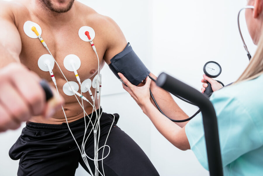
- Exercise Stress Test (Treadmill Stress Test): In this version of the test, the patient walks or runs on a treadmill while connected to an electrocardiogram (ECG) machine, which records the heart’s electrical activity. As the patient exercises, the workload on the heart increases, and if there are any blood flow abnormalities due to CAD, they become more evident. The patient’s heart rate, blood pressure, and symptoms are closely monitored during the test.
- Pharmacological Stress Test: In cases where a patient is unable to exercise due to physical limitations or when an exercise stress test is inconclusive, pharmacological stress agents are used to induce the same effects on the heart as exercise. These agents, such as adenosine, dipyridamole, or dobutamine, dilate the coronary arteries, simulating the stress experienced during exercise.
Preparing for the Test
Patients are usually asked to fast for a certain period before the test. Medications might need to be adjusted, and it’s important to avoid caffeine. Comfortable clothes and shoes suitable for exercise should be worn.
The Nuclear Stress Test Procedure:
The nuclear stress test is generally performed in a specialized cardiology or nuclear medicine department by a team of trained medical professionals, including cardiologists, nuclear medicine technologists, and nurses. The procedure typically involves the following steps:
1. Pre-Test Preparations:
Before the nuclear stress test, the patient’s medical history, symptoms, and current medications are reviewed by the healthcare team. Some medications that could interfere with the test may need to be temporarily stopped. The patient is instructed not to eat or drink anything containing caffeine for at least 24 hours before the test, as caffeine can affect the test results.
2. Placement of Intravenous (IV) Line:
At the beginning of the test, an IV line is inserted into the patient’s arm. This allows for the injection of a small amount of radioactive tracer, which is essential for nuclear imaging.
3. Resting Phase (Pre-Stress Imaging):
During the resting phase, the patient is asked to lie down on an examination table, and a small amount of the radioactive tracer is injected through the IV line. The tracer travels through the bloodstream and is taken up by the heart muscle, highlighting areas of healthy blood flow. A gamma camera, a specialized imaging device, is positioned over the chest to capture images of the heart at rest.
4. Stress Phase (Exercise or Pharmacological Stress):
If the patient is undergoing an exercise stress test, they will be asked to walk or run on a treadmill while the speed and incline gradually increase. The goal is to reach the target heart rate based on the patient’s age and physical condition. During this phase, the ECG, blood pressure, and symptoms are continuously monitored.
If a pharmacological stress test is being performed, the stress-inducing medication is administered through the IV line, and the patient’s heart rate and blood pressure are closely monitored.
5. Post-Stress Imaging:
After the stress phase, the patient is quickly moved back to the imaging table, and a second set of images is obtained using the gamma camera. These images show the heart’s blood flow during stress conditions, allowing the healthcare team to compare it with the resting images.
6. Image Analysis and Interpretation:
The collected images are then carefully analyzed by experienced nuclear medicine technologists and interpreted by cardiologists. They assess the distribution of the radioactive tracer in the heart, identifying areas with reduced blood flow (also known as “cold spots”) that may indicate CAD or other cardiac abnormalities.
7. Post-Test Monitoring and Follow-Up:
Once the test is complete, the IV line is removed, and the patient is allowed to rest. In most cases, patients can resume their regular activities after the test. The results are discussed with the patient during a follow-up appointment, where the healthcare provider will explain the findings and recommend further steps if necessary.
Safety Considerations of the Nuclear Stress Test:
The amount of radioactive tracer used in the nuclear stress test is minimal and considered safe. The radiation exposure from the test is comparable to other diagnostic imaging procedures, such as a CT scan or X-ray. The benefits of obtaining critical cardiac information through this test outweigh the minimal risks associated with radiation.
However, certain patients, such as pregnant women and individuals with severe kidney problems, may not be suitable candidates for a nuclear stress test due to potential risks. It is essential for patients to disclose their medical history and any existing conditions to the healthcare team before undergoing the test.
Interpreting the Results
A cardiologist compares the images from the stress and resting scans. If areas of the heart show less tracer uptake during the stress scan compared to the resting scan, it may indicate reduced blood flow to the heart muscle due to blocked or narrowed arteries.
The nuclear stress test is a powerful diagnostic tool that plays a pivotal role in the field of cardiology. By evaluating the heart’s function and blood flow under stress conditions, it enables early detection and accurate diagnosis of coronary artery disease. Through the information obtained from this test, healthcare providers can tailor treatment plans, monitor disease progression, and make informed decisions about the best course of action for each patient. The non-invasive nature and accuracy of the nuclear stress test make it an invaluable procedure in safeguarding heart health and promoting better cardiovascular outcomes for patients worldwide. As medical technology continues to advance, we can expect further refinements to this procedure, offering even greater precision and effectiveness in detecting and managing cardiac conditions.


