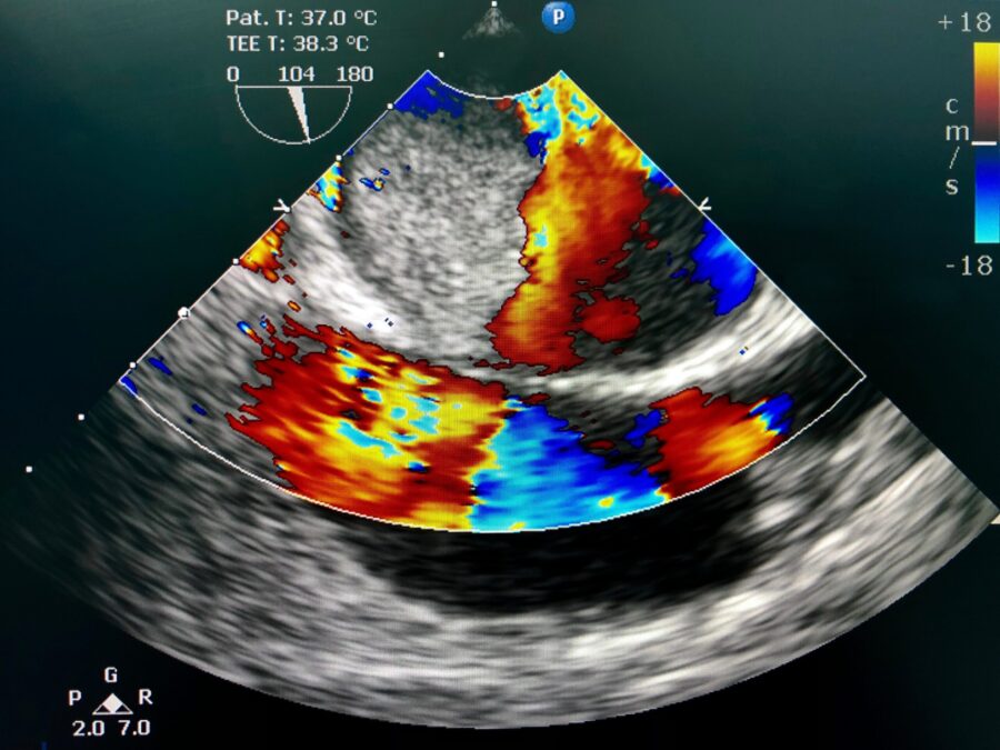
Transesophageal echocardiogram (TEE) is a powerful diagnostic tool in the cardiology field, allowing doctors to get a closer and more detailed look at the heart. This procedure provides invaluable information, guiding the diagnosis and management of various heart conditions. In this comprehensive guide, we delve into the details of TEE, its benefits, and what patients can expect during the process.
Understanding Transesophageal Echocardiogram (TEE)
A TEE is a type of echocardiogram where the ultrasound probe is inserted into the esophagus, which lies just behind the heart. This positioning provides an unobstructed, close-up view of the heart, leading to clearer, more detailed images compared to a traditional echocardiogram.
Why Doctors Recommend Transesophageal Echocardiogram (TEE)
A TEE is often recommended when:
- A traditional echocardiogram doesn’t provide sufficient information.
- The doctor needs a closer look at the heart valves or the upper chambers of the heart.
- The presence of blood clots, tumors, or infectious conditions is suspected.
The Transesophageal Echocardiogram (TEE) Procedure
While specifics may vary, the following steps are commonly involved in a TEE procedure:
- The patient’s throat is numbed, and mild sedation is usually provided.
- The ultrasound probe is gently guided down the patient’s esophagus.
- Images of the heart are then captured for analysis.
Preparing for a TEE
Proper preparation is crucial for the success of a TEE procedure. Patients are usually asked to fast for at least six hours before the procedure. They should also discuss all medications they’re currently taking with their doctor, as some might need to be adjusted or temporarily stopped. And since sedation is involved, patients need to arrange for a ride home post-procedure.
Understanding the TEE Report
TEE reports provide a detailed analysis of the heart’s structure and function. They may include information on the size and function of the chambers, the condition of the valves, and the presence of any abnormal masses or clots. Understanding these technical details can be challenging for patients, but their healthcare provider can help interpret the results and explain what they mean in terms of diagnosis or treatment.
A TEE is an invaluable tool that aids doctors in diagnosing and managing many heart conditions. By understanding what to expect from the procedure, patients can feel more comfortable and prepared on the day of the exam. With its detailed imagery and minimally invasive nature, TEE has revolutionized cardiac care, allowing for precise diagnoses and treatment planning, thus contributing to better patient outcomes.
Advancements in TEE Technology
The technology behind TEE has evolved significantly over the past few decades. The introduction of three-dimensional (3D) imaging has been a game-changer, providing more detailed and realistic views of the heart’s structures. 3D-TEE has become instrumental in preoperative planning for heart surgeries, especially valve repairs or replacements, as it allows surgeons to visualize the precise nature and extent of the pathology.
Intracardiac echocardiography (ICE) is another advancement that combines TEE with cardiac catheterization procedures. It offers real-time imaging of the heart’s interior during procedures like ablation or device implantation, improving accuracy and safety.
The Role of TEE in Guiding Treatments
TEE is not just a diagnostic tool; it’s an invaluable guide for numerous interventional cardiology procedures. It can assist in navigating catheters during complex procedures such as left atrial appendage closure, percutaneous valve replacements, or septal defect closures.
TEE also plays an essential role during surgical procedures. For instance, intraoperative TEE is routinely used in open-heart surgeries to guide surgeons and assess the success of the procedure immediately.
TEE versus Other Imaging Modalities
Compared to other cardiac imaging techniques like a standard echocardiogram (transthoracic echocardiogram, TTE) or cardiac MRI, TEE offers a few distinct advantages. As it bypasses the interference of the lungs and chest wall, it provides superior image resolution. This allows for better evaluation of certain structures like the left atrial appendage, pulmonary veins, or the aorta.
However, every imaging technique has its strengths and weaknesses, and the choice often depends on the patient’s condition, the clinical question at hand, and local expertise and resources.
Common Misconceptions About TEE
It’s worth addressing some common misconceptions about TEE. First, while TEE involves the insertion of a probe down the esophagus, it is generally well-tolerated due to the sedatives and throat-numbing agents used. The procedure is not painful, though some mild discomfort or gagging sensation might be experienced.
Second, the risk of serious complications, like esophageal perforation, is exceedingly low. The procedure is considered quite safe when performed by experienced professionals in a controlled setting.
The Future of TEE
The field of echocardiography is continuously evolving, and the future of TEE looks promising. Advancements in technology will likely lead to higher-resolution images and even more detailed views of heart structures. Innovations in 3D imaging and the integration of artificial intelligence may allow for automated measurements and more sophisticated analyses.
In conclusion, TEE is a significant advancement in cardiac imaging, providing detailed information that can guide diagnosis and treatment. The more patients understand about this procedure, the better prepared they will be to navigate their healthcare journey, from preparation to recovery and understanding the results. It’s another tool in the toolkit of cardiac care that, when combined with the healthcare team’s expertise, can help ensure the best outcomes for patients.


