In the medical world, one of the most detailed and efficient ways to image the heart and surrounding structures is through Cardiac Magnetic Resonance Imaging (Cardiac MRI). This non-invasive test creates detailed pictures of the heart’s structure and function, aiding in the diagnosis and treatment of heart conditions. This comprehensive guide will delve into the specifics of a cardiac MRI, its purpose, its process, and the interpretation of its results.
Understanding Cardiac MRI
A cardiac MRI is a specialized type of MRI tailored to capture detailed images of the heart. Like other MRI scans, a cardiac MRI uses a strong magnetic field and radio waves to generate pictures of the heart. The imaging can depict the heart’s chambers, blood vessels, valves, and overall structure in unparalleled detail. Unlike X-rays and CT scans, MRIs do not use ionizing radiation, making them a safe choice for most patients.
Why Doctors Recommend Cardiac MRI
A cardiac MRI is an exceptional tool in a doctor’s diagnostic repertoire for many reasons. Primarily, it is used when other tests, like echocardiograms or cardiac CT scans, are not sufficient or cannot be used due to specific patient conditions. Cardiac MRIs offer a more detailed, cross-sectional view of the heart, which can help doctors diagnose a variety of cardiovascular conditions, including:
- Congenital heart defects
- Coronary heart disease
- Damage from heart attacks
- Heart failure
- Heart valve problems
- Pericarditis (inflammation of the lining around the heart)
- Cardiac tumors
Moreover, cardiac MRIs are useful for guiding the recovery plan after heart surgery or as part of a strategy for managing ongoing heart conditions.
The Cardiac MRI Procedure
A cardiac MRI is typically an outpatient procedure performed in a hospital or specialized imaging center. Patients are asked to wear a hospital gown and remove any metal objects, including jewelry and watches, as they can interfere with the magnetic field.
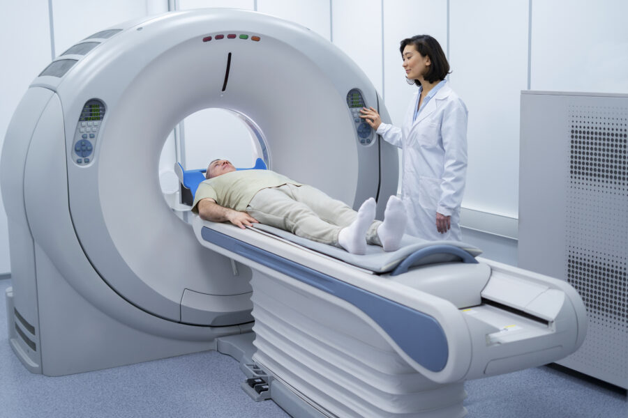
During the procedure, the patient lies on a sliding table that goes inside a large cylindrical machine – the MRI scanner. An intravenous line (IV) is usually inserted into a vein in the patient’s arm to administer a contrast agent, which improves the clarity of the images by highlighting the heart and blood vessels. The contrast agent is usually gadolinium, which has a very low risk of causing allergic reactions.
The MRI machine produces a strong magnetic field around the patient, and radio waves are sent to the body, which responds by sending back signals. These signals are collected and processed by a computer to create detailed images of the heart from various angles.
The test usually lasts between 45 minutes to an hour and a half, depending on what information the doctor needs. The patient must lie very still during the procedure and may be asked to hold their breath at times to prevent blurring of the images.
Life After a Cardiac MRI
After a cardiac MRI, patients can usually resume their normal activities immediately. If a contrast agent was used, it will naturally leave the body through the kidneys over time. Patients are usually advised to drink plenty of water to help eliminate the contrast agent from the body.
Interpreting the Results
The results of a cardiac MRI are analyzed by a radiologist – a doctor specialized in interpreting medical images. The radiologist will examine the images to assess the structure and function of the heart, looking for any signs of damage, disease, or anomalies. A detailed report is then prepared and sent to the patient’s doctor.
Based on the findings of the cardiac MRI, the doctor may recommend further testing or specific treatments. The images can also be used to guide surgical procedures or assess the effectiveness of ongoing treatment. Importantly, results should be discussed thoroughly with the healthcare provider, who can provide context and explain the implications of the findings.
In conclusion, a cardiac MRI is a highly detailed, non-invasive, and safe imaging technique that plays a pivotal role in diagnosing and managing heart conditions. Its ability to create comprehensive images of the heart’s structure and function makes it an invaluable tool in cardiovascular care. By understanding the purpose and process of a cardiac MRI, patients can approach the procedure with greater confidence and peace of mind. As with any medical procedure, all questions and concerns should be addressed with the healthcare provider to ensure clarity and comfort with the process.


